The progression of mitosis illustrated
I compiled the following set of microscopy images to teach an undergraduate student how to recognize mitotic phases.
They represent fixed HeLa cells whose DNA has been stained with Hoechst; the cells express a GFP-CENP-A fusion protein, which localizes to the centromeres (giving the green dots staining).
A PDF version (one A4 page) of this note is also available.
|
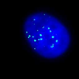
Interphase. |
|
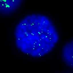
Early prophase; condensation of chromosomes begins. |
|
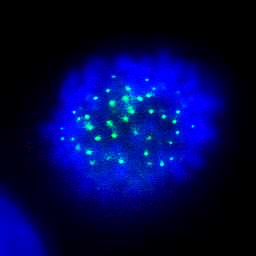
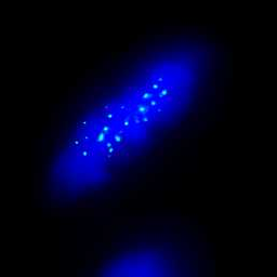
Late prophase: chromosomes are fully condensed and form a circle in the (disappearing) nucleus. |
|
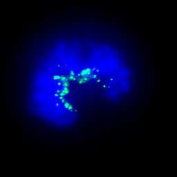
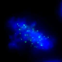
Prometaphase; the circle opens as chromosomes are mobilized to form the metaphase plate (congression). |
|
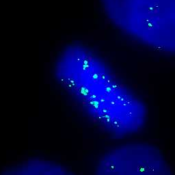
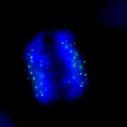
Metaphase; chromosomes are aligned on the metaphase plate. |
|
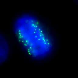
Abnormal metaphase; one or several chromosomes failed to align in the metaphase plate. |
|
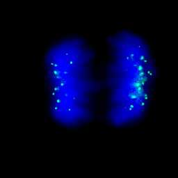
Metaphase-to-anaphase transition (late metaphase); the distance between centromeres increases. |
|
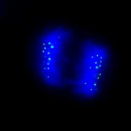
Early anaphase; chromosomes start moving towards the poles (segregation). |
|
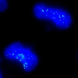
Mid-anaphase; chromosomes now form two distinct shapes. |
|
Late anaphase; the gap between the two sets of chromosomes widens. |
|
Abnormal anaphase; presence of lagging chromosomes (or chromosome fragments) between the two sets of migrating chromosomes. |
|
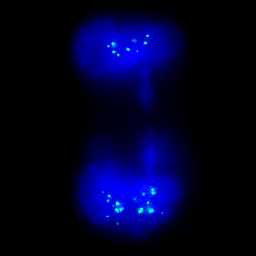
Telophase; the two chromatin shapes become larger as the chromosomes begin to decondensate. |
|
Abnormal telophase; presence of a “chromatin bridge” (partial or completeà between the two sets of chromosomes. |
|
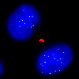
Cytokinesis; chromosomes are decondensed; chromosomal passenger proteins (red, Aurora B kinase) concentrate on the midbody between the two newly formed nuclei. |